
Measure the lesion Review the result
SKInguide+ MEASURE
Objective image processing, with clinician correction, annotation and reporting
Turn an image into structured information
INSIDE MEASURE
Explore the measurements in the interface. Select an output to see where it appears and what it means.
From image capture to a clinician-reviewed report.
HOW MEASURE WORKS
Connect the image to the case, review the measurements, and bring the clinician’s findings into the record.
01 / CAPTURE
Link and capture
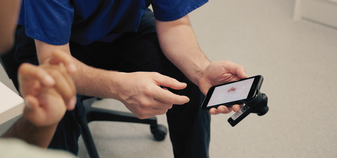
Open the case and connect the smartphone using its QR code. Capture the dermoscopic image and check the image quality before continuing.
02 / MEASURE
Check and refine
Generate the outline and measurements. Review the border, correct it where needed, and apply the update.
03 / DOCUMENT
Review and approve
Add clinical findings and the clinician’s diagnosis. Review the draft report before saving and exporting the approved information.
CONTROL STAYS WITH THE CLINICIAN
A starting point you can correct
The proposed border is available for review. If it needs adjusting, the clinician can place points around the lesion or begin a manual outline, then apply the changes.
Diameter, area, and colour results are updated based on the reviewed outline.
PROPOSE
Generate the measurements
Inspect the outline and its associated results.
REFINE
Apply your correction
Review the updated measurements before documentation.
CHOOSE YOUR LEVEL OF SUPPORT
Measure the image Add Insight when needed
MEASURE
Image-quality checks, border review, measurements, and clinical documentation. No AI diagnostic decision support.
INSIGHT
Everything in Measure, with AI features, uncertainty, and decision support for clinician review.
DEVELOPMENT
A working interface Evaluation ahead
SkinGuide+ remains in development. Prospective clinical evaluation and EMIS integration are development priorities.
-
The example shows calibrated physical measurements. A valid image scale is needed to convert pixels into millimetres or square millimetres.
-
No. Measure provides image measurements and documentation tools. The clinician records their own assessment.
-
Yes. The interface provides tools to correct the outline or start a manual outline, then apply the change to update measurements.